


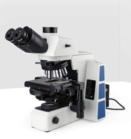
After years of research and development in optical technology field, XJE500 biological microscope is designed to present a safe, comfortable and efficiency observation experience for users. With perfectly performed structure, high-definition optical image and simple operating system, XJE500 realizes professional analysis, and meets all the needs of research in scientific, medical and other fields.
1. Introduction
• High eye-point ultra wide field plan eyepieces
Breaking through 22mm conventional field of view, reaching to 25mm and 26.5mm,much smoother and wider field of view, is helpful to improve the work efficiency.
Locating pin on eyepiece inserts into the eyepiece tube, fixing the eyepiece for easy focusing.Larger adjustable range of diopter from -8 to +5, meets more demands of different users.
Eyepiece cup can be turn up to avoid external stray light. Spectacle-wearers should turn down the cup to protect both spectacles and eyepieces.
• Large stage with adjustment in either hand
In order to correct the shortcoming of horizon guide rail, new stage is designed with double-way linear driving mechanism. This change protects the stage from overload at the end of both rails, improves the rigidity and performance of the stage.
• Modular frame, improving the system compatibility
XJE500 with modularization design, separated cross arm and main body, improves the system compatibility of biological and fluorescence frame.
• Highly sensitive coaxial coarse and fine adjustment
Coaxial adjustment adopts double-stage driving, with adjustable tension tightness and upper limit stop, coarse range is 25mm and fine precision is 1μm. Not only accurately focus but also precision measurement is available.
2. Structure


3. Image
1) Brightfield observation:

Adenoma of colon Endometrial cancer
2) Phase contrast observation:

Human oral epithelial cells Cheek epithelial cell
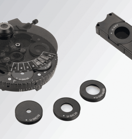
3) DIC observation:

Human oral epithelial cells Bovine pulmonary artery endothelial cells